Detailed Guideline on Pyroptosis Research
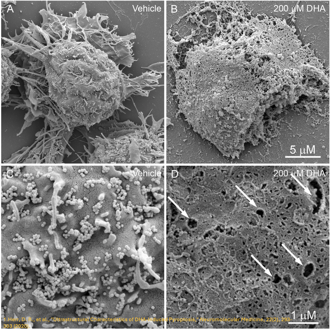
 Pyroptosis research involves several key experimental techniques to study this form of programmed cell death. Scanning electron microscopy (SEM) provides detailed observations of the characteristic membrane rupture and morphological changes that occur during pyroptosis. Western blot (WB) can be used to detect pyroptosis-related proteins such as gasdermin D and inflammasome components. Enzyme-linked immunosorbent assay (ELISA) helps quantify inflammatory cytokines like IL-1β and IL-18, which are crucial markers of pyroptosis. Immunofluorescence (IF) microscopy is useful for visualizing cellular localization of pyroptotic markers. These methods together offer a comprehensive understanding of pyroptosis mechanisms and their regulation.
Cell Morphology Detection
1. Scanning Electron Microscopy (SEM) for Observing Cell Morphology
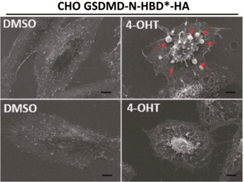
Figure 1. Scanning Electron Microscopy Observations of Pyroptotic Cell Morphology [1]
Representative SEM images of CHO cells expressing GSDMD-N-HBD*-HA, treated with DMSO or 4-OHT for 1 hour, are shown. Arrows highlight the bubbling characteristic of pyroptotic cells. SEM analysis showed that the dying cells stayed attached to the culture surface and released multiple pyroptotic bodies.
2. Immunofluorescence Staining
 Figure 2. Immunofluorescence Staining of GSDMD [1]
Deconvolution microscopy was performed on CHO cells expressing various GSDMD-N-HBD*-HA deletion mutants, following a 1-hour treatment with 4-OHT. After treatment, cells were immunostained for HA and counterstained with Hoechst and PI (Scale bar: 5 μm). PI-positive cells were considered dead. Immunostaining results showed that GSDMD-N-HBD*-HA deletion mutants DN1 to DN4 exhibited behavior similar to wild-type GSDMD-N-HBD*-HA.
Cell Detection of Pyroptosis-Related Proteins
The biochemical characteristics of pyroptosis are primarily marked by the formation of inflammasomes, activation of caspases and gasdermins, and the release of numerous pro-inflammatory cytokines.
1. Western Blot Method for Detecting the Expression Levels of Pyroptosis-Related ProteinsDuring pyroptosis, Gasdermin D (GSDMD, 53 kD) is cleaved to produce an active fragment of approximately 30 kD. Similarly, pyroptotic activation involves the cleavage and activation of Caspase-1, Caspase-4, and Caspase-5, leading to the processing of pro-inflammatory cytokines (e.g., IL-1β, IL-18) and the induction of pyroptotic cell death. The activity of these caspases can be confirmed by detecting their enzymatic cleavage products, while the expression levels of pyroptosis-related proteins can be assessed through Western Blot (WB) analysis.

Figure 3. WB Detection of Pyroptosis-Related Targets [2]
Caspase-3 activation and GSDME processing were reduced in CASP1/CASP8 double knockout (DKO) cells compared to CASP1 knockout (KO) cells. These findings indicate that caspase-8 plays a key role in initiating GSDME processing during caspase-1-independent pyroptosis.
ABclonal Pyroptosis-Related Target Antibody Product Recommendations:
|
Category |
Research Target |
Product Number |
Product Name |
Application |
Species |
|
Inflammasome Components (Sensors) |
NLRP3 |
NLRP3 Rabbit pAb |
WB, IF/ICC, ELISA |
Human, Mouse, Rat |
|
|
NLRP3 Rabbit pAb |
WB, IF/ICC, ELISA |
Human, Mouse, Rat |
|||
|
NLRC4 |
NLRC4 Rabbit pAb |
WB, IF/ICC, ELISA |
Human, Mouse, Rat |
||
|
NLRP6 |
NLRP6 Rabbit pAb |
WB, ELISA |
Human, Mouse, Rat |
||
|
Inflammasome Components (Adapter Molecules) |
ASC / TMS1 |
ASC/TMS1 Rabbit pAb |
WB, IHC-P, IF/ICC, ELISA |
Human, Mouse, Rat |
|
|
ASC/TMS1 Rabbit pAb |
WB, IHC-P, IF/ICC, IP, ELISA |
Human, Mouse, Rat |
|||
|
ASC/TMS1 Rabbit mAb |
WB, IHC-P, ELISA |
Human, Mouse, Rat |
|||
|
Inflammasome Components (Pro-inflammatory Caspases) |
Caspase-1 |
Caspase-1 Rabbit pAb |
WB, IF/ICC, ELISA |
Human, Mouse, Rat |
|
|
Caspase-1 Rat mAb |
WB, ELISA |
Mouse, Rat |
|||
|
Caspase-4 |
Caspase-4 Rabbit pAb |
WB, IHC-P, ELISA |
Human |
||
|
Pro-inflammatory Cytokines |
IL1β |
IL1β Rabbit mAb |
WB, IF/ICC, ELISA |
Human |
|
|
IL18 |
IL18 Rabbit pAb |
WB, IP, ELISA |
Human |
||
|
IL18 Rabbit mAb |
WB, IHC-P, ELISA |
Human, Mouse, Rat |
|||
|
Pyroptosis Effector Molecules |
GSDMD |
GSDMD (Full length+C terminal) Rabbit mAb |
WB, ELISA |
Human |
|
|
GSDMD (Full length+C terminal) Rabbit pAb |
WB, IF/ICC, ELISA |
Human, Mouse, Rat |
|||
|
DFNA5/GSDME |
GSDME (Full length+N terminal) Rabbit pAb |
WB, IHC-P, IF/ICC, ELISA |
Human, Mouse, Rat |
2. ELISA Detection of IL-1β, IL-18, and Other Inflammatory Cytokines
The inflammatory cytokines IL-1β and IL-18 are activated and secreted in a caspase-1-dependent manner during pyroptosis. Interleukin-1β (IL-1β) is a potent endogenous pyrogen that can induce fever, leukocyte migration to tissues, and the expression of various cytokines and chemokines. Interleukin-18 (IL-18) promotes the production of interferon-γ and is essential for the activation of T cells, macrophages, and other immune cells. Both IL-1β and IL-18 play critical roles in the pathogenesis of various inflammatory and autoimmune diseases.

Figure 4. Detection of IL-1β levels in the supernatant using the ELISA method [2]
In contrast to IL-1α release, IL-1β release was inhibited in macrophages treated with VX765 or MCC950.
ABclonal Pyroptosis-Related Target ELISA Kits Recommendations:
|
Product Code |
Product Name |
Application |
Species |
|
Human IL-1 beta ELISA Kit |
ELISA |
Human |
|
|
Mouse IL-1 beta ELISA Kit |
ELISA |
Mouse |
|
|
Mouse IL-1 beta FAST ELISA Kit |
ELISA |
Mouse |
|
|
Rat IL-1 beta ELISA Kit |
ELISA |
Rat |
|
|
Mouse IL-18 ELISA Kit |
ELISA |
Mouse |
|
|
Human IL-18 ELISA Kit |
ELISA |
Human |
Recommended Representative Literature on Pyroptosis Research:
- Chen et al., Pyroptosis is driven by non-selective gasdermin-D pore and its morphology is different from MLKL channel-mediated necroptosis. Cell Res 26, 1007-1020 (2016).
- Aizawa et al., GSDME-Dependent Incomplete Pyroptosis Permits Selective IL-1alpha Release under Caspase-1 Inhibition. iScience 23, 101070 (2020).


