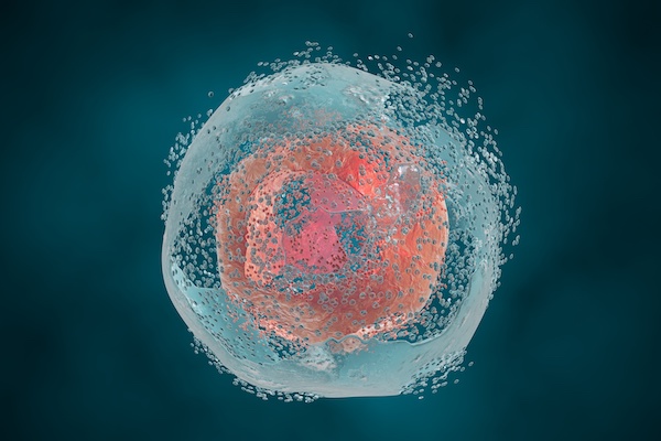
In the 1980s and 1990s, scientists discovered that macrophages exposed to bacterial toxins or infections underwent a unique form of cell death that required the activation of caspase-1. For years, this process was mistakenly classified as apoptosis, a non-inflammatory and orderly form of cell death. However, unlike apoptosis, this newly observed cell death was marked by swelling, rupture, and the release of inflammatory signals, indicating a far more chaotic and immune-activating process. It wasn’t until 2001 that Cookson BT and Brennan MA officially named this inflammatory form of cell death “pyroptosis”. [2] Since then, research has revealed that pyroptosis plays a key role in immune defense by promoting inflammation in response to infections. The discovery of gasdermin D (GSDMD), a protein that forms pores in the cell membrane, further clarified the mechanism of pyroptosis. Today, it’s understood that pyroptosis occurs in various cell types and is critical in both fighting infections and contributing to inflammatory diseases when dysregulated.
What is Pyroptosis?
Pyroptosis is a type of programmed cell death triggered by inflammasomes. It is characterized by the cell swelling until its membrane ruptures, leading to the release of cell contents and a strong inflammatory response. The occurrence of pyroptosis depends on inflammatory caspases and the Gasdermin (GSDMs) protein family, accompanied by the release of numerous pro-inflammatory factors. Pyroptosis is a crucial component of the body's innate immune response and plays an important role in combating infections.

Figure 1. Core Molecular Mechanisms of Autophagy, Pyroptosis, Ferroptosis, and Necroptosis [3]
Cell Pyroptosis Signaling Pathway
Pyroptosis is an inflammatory form of cell death that relies on the activation of certain members of the caspase family by inflammasomes. This activation cleaves and activates gasdermin proteins. The activated gasdermin proteins then translocate to the cell membrane and form pores. Due to the difference in osmotic pressure between the inside and outside of the cell, the cell continuously swells until it eventually ruptures, releasing its contents and triggering a strong inflammatory response.
Classical and Non-Classical Pathways of Pyroptosis
1. Classical Pathway (Caspase-1 Dependent Pathway)Caspase-1-dependent pyroptosis relies on the activation of canonical inflammasomes. In this pathway, pathogen-associated or danger-associated molecular patterns engage their respective inflammasome sensors, such as NLRP1b, NLRP3, NLRC4, AIM2, or Pyrin. As shown in Figure 2, the NLRP3 and NLRC4 inflammasomes require the kinase NEK7 and ligand-binding NAIP proteins for activation, respectively. Once activated, inflammasome sensors recruit the adaptor ASC and the cysteine protease caspase-1 into a macromolecular complex. Caspase-1 then cleaves gasdermin D and the precursor cytokines pro-IL-1β and pro-IL-18, triggering pyroptosis and the maturation of IL-1β and IL-18. The 31-kDa N-terminal fragment of gasdermin D forms pores in the host cell membrane, facilitating the release of cytoplasmic contents. [4]
Note: An inflammasome is a multi-protein signaling complex that assembles in the cytoplasm upon detection of danger signals from the host or pathogens. This assembly promotes the release of cytokines, pyroptosis, and inflammation. The known inflammasomes include NLRP1 inflammasome, NLRP3 inflammasome, NLRC4 inflammasome, IPAF inflammasome, and AIM2 inflammasome.
2. Non-canonical Pathway (caspase-4, 5, 11-dependent pathway)The non-canonical pathway, also known as the caspase-1-independent pathway or non-classical inflammasome signaling pathway, is mediated by human caspase-4 and caspase-5, or mouse caspase-11. These inflammatory caspases can directly cleave gasdermin D and initiate pyroptosis. The N-terminal fragment of gasdermin D, after cleavage, can also activate the NLRP3 inflammasome, leading to the caspase-1-dependent maturation of IL-1β and IL-18.

Figure 2. Molecular mechanisms of caspase-1-dependent and -independent pyroptosis [4]
The two pathways mentioned above are the earliest deciphered cell pyroptosis pathways. In recent years, as research has deepened, the definition and related mechanisms of cell pyroptosis have continued to be revised and refined.
3. Incomplete PyroptosisIn macrophages lacking Caspase-1/11, the inflammasome NLRP3 can activate Caspase-8, which in turn cleaves GSDME, leading to a form of incomplete pyroptosis. It is considered incomplete because this process does not involve the release of the key pyroptosis factor IL-1β, but rather the secretion of IL-1α.

Figure 3. GSDME-dependent incomplete cell pyroptosis allows selective release of IL-1α under Caspase- 1 inhibition [5].
After 2015, the mechanisms of pyroptosis mediated by gasdermin family members have gradually been refined. Related research has confirmed that the ultimate effector proteins of pyroptosis are members of the gasdermin family. Pyroptosis is now redefined as "gasdermin-mediated programmed necrotic cell death." Among the gasdermin family, Gasdermin D (GSDMD) is a common substrate for both caspase-1 and caspase-4/5/11. Caspase-1 and caspase-4/5/11 can cleave GSDMD [6]. GSDMD is composed of an N-terminal domain (GSDMD-N) and a C-terminal domain (GSDMD-C). By releasing the cleaved GSDMD N-terminal domain, GSDMD-N possesses intrinsic pore-forming activity. In its full-length state, GSDMD-C can inhibit the activity of GSDMD-N. Once GSDMD is cleaved and activated, the released GSDMD-N can bind to membrane lipids and form pores, leading to the release of cytokines and various cytoplasmic contents, ultimately causing cell membrane rupture and pyroptosis[7]. GSDME can be specifically cleaved and activated by caspase-3, leading to pyroptosis[8]. Granzyme A (GZMA) secreted by cytotoxic lymphocytes cleaves GSDMB to release the GSDMB-N fragment, which induces cell perforation and triggers pyroptosis[9].
For more information, visit our Pyroptosis Learning Center .
Recommended Representative Literature on Pyroptosis Research:
-
- Herr, D. R., et al., "Ultrastructural Characteristics of DHA-Induced Pyroptosis," Neuromolecular Medicine, 22(2), 293-303 (2020).
- Cookson, B. T., et al., "Pro-inflammatory Programmed Cell Death," Trends in Microbiology, 9(3), 113-114 (2001).
- Gao, X., et al., "Autophagy, ferroptosis, pyroptosis, and necroptosis in tumor immunotherapy," Signal Transduction and Targeted Therapy, 7, 196 (2022).
- Man, M., et al., "Molecular mechanisms and functions of pyroptosis, inflammatory caspases, and inflammasomes in infectious diseases," Immunological Reviews, 277, 61-75 (2017).
- Aizawa, Y., et al., "GSDME-Dependent Incomplete Pyroptosis Permits Selective IL-1α Release under Caspase-1 Inhibition," iScience, 23, 101070 (2020).
- Shi, J., et al., "Cleavage of GSDMD by inflammatory caspases determines pyroptotic cell death," Nature, 526, 660-665 (2015).
- Liu, Z., et al., "Structures of the Gasdermin D C-Terminal Domains Reveal Mechanisms of Autoinhibition," Structure, 26(5), 778-784.e3 (2018).
- Wang, Y., et al., "Chemotherapy drugs induce pyroptosis through caspase-3 cleavage of a gasdermin," Nature, 547, 99-103 (2017).
- Zhou, Z., et al., "Granzyme A from cytotoxic lymphocytes cleaves GSDMB to trigger pyroptosis in target cells," Science, 368 (2020).


