 Having worked in a proteomics lab for my PhD dissertation, I had some familiarity with the tools and strategies used to study biology on a systems level. One of the concepts I was always interested in was the ability to just follow a protein's journey throughout the cell, from the time it is translated by the ribosome to its final destination either within an organelle or when it is secreted into the extracellular space. At the time I was finishing up, I wasn't sure that the technology was yet advanced enough to make that a reality, particularly if done within a single cell. But within the past few years, a new era of spatial proteomics has emerged to allow us to observe cell biology in a whole new light.
Location is Key
While I was in graduate school, my proteomics work was based on the quantity and modifications of proteins within a cell population, so this would be samples representative of hundreds of thousands to millions of cells of a single type. The proteome consists of the totality of proteins present in the cell type, tissue, or body fluid of interest, and lends a wider view of biology at the systems level. Of course, you can compare the proteome of two different cell lines, for example MCF-7 against a fibroblast line, but the information you get from that, while still important, is lacking the fine details. For instance, where are the proteins localized to in a breast cancer cell line compared to a leukemia cell line? In an organism, can you tell where a protein of interest is within a liver cell versus a brain cell? Traditional techniques can tell you if that protein exists within the tissue, but to simultaneously quantify abundance and localization was a challenge due to the limitations of technology at that time.
 Because protein localization is key to its functions, both within the cell and the organism as a whole, it is important to note the precise localization of the target protein(s) to better understand their role in human health and disease, which will contribute to a better understanding of cell biology. Over the years, as microscopes and mass spectrometers have gotten better to provide greater resolution, the field of spatial proteomics has benefited from these technological advances. 1 As the field becomes more mainstream, we can imagine a world where spatial proteomics can go hand in hand with traditional cell biology and biomedical research, creating analytical tools that will allow the greater use of vast data sets in understanding cellular processes and disease states.
Principles of Spatial Proteomics
Proteomics has revealed a complexity that includes cell-to-cell variations in protein dynamics, including their localization to specific intracellular compartments. Comparing the spatial proteomics between cells could reveal differences between a "healthy" cell versus an "aberrant" cell and could potentially serve as a discovery or diagnostic tool to further lock in to understanding the functions of those proteins of interest as well as to fine tune treatment strategies. 1
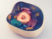
As technology improves and the amount of input material required decreases, the increased resolution and utility allows scientists to do more with less, and that includes applying those samples to spatial proteomic analysis. As the proponents of spatial proteomics attest to, subcellular localization of the constituents of the proteome can affect the function of those proteins, due to differences (both minute and significant) in the local protein and macromolecule content as well as the buffer system within their compartment. The initial use of spatial proteomics was to determine the proteomes within major organelles, but with ever-advancing imaging systems, scientists have learned that there are many unique subcellular compartments, membrane-bound or just sequestered into specific regions of the cytosol, and many proteins are found in several different compartments within a single cell. This complexity can be more effectively dissected now with high-throughput imaging as well as higher-resolution mass spectrometry to simultaneously query the localization and the abundance of proteins within their target compartments.
 Studying biology, including proteomics, on a single-cell level is highly attractive because the fine details of biological function are no longer lost within a population, not to mention the desire to reduce resources and labor in generating samples at volume. To this end, scientists and facilities such as the Pacific Northwest National Laboratory have innovated new platforms to study spatial proteomics on single cells. PNNL's platform is known as Nanodroplet Processing in One Pot for Trace Samples, or nanoPOTS, and is capable of downscaling sample processing volumes to under 200 nL. Some of the applications boasted by PNNL include tracking the development of sensory hair cells, profiling proteins within single circulating tumor cells from blood samples. The multidisciplinary and multitechnology nature of spatial proteomics also allows for the development of automated platforms, including microdissection tools to study tissue proteomes, and the use of artificial intelligence and machine learning to parse the large datasets, similarly to what has already been done for genomic data and neurobiology research. 2, 3
Applications of Spatial Proteomics
 As suggested by Georg Borner, one of the many pioneers of spatial proteomics, many human disorders are associated with defective protein transport (you can read more about vesicular transport here), which means the more comprehensive mapping of protein localization events could provide greater insight into both cell biology and disease progression. There are myriad experimental and medical applications that could be yielded from spatial proteomics, particularly in pathology and diagnostics. 3 The list is immense, and includes cell-cell and protein-protein interactions and biomarker expression. The integration of automated platforms and enhanced analysis with the aid of machine learning can help scientists and pathologists study diverse physiological phenomena such as the immune infiltration within tumor microenvironments, aortic plaques in a human heart, and even aid in neurobiology such as brain injury models and Alzheimer's disease. 3, 4 The marriage of human ingenuity, technological advances, and optimized analytical strategies suggest a near-limitless potential for this developing field!
Shining a Light On Signal Transduction
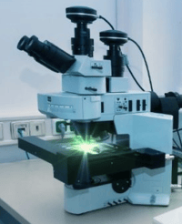 Recently, MIT researcher and Howard Hughes Medical Institute investigator Edward Boyden and his group reported a new approach to image the activity inside a cell. Published open access in Cell,5 this methodology employs fluorophores that exhibit distinct clocklike properties that can be used to track different cellular signaling molecules. Once a cell is "triggered," a recording of the fluorescence microscopy data is produced over a period of a few seconds and the imaging data can be parsed into signaling amplitudes that give a more comprehensive picture of the intensity and the movement of those signaling molecules being studied. Termed temporally multiplexed imaging, or TMI (which probably does give "too much information" but in biology you can never have too much data), the methodology only requires a standard fluorescence microscope and genetically encoded fluorescent reporters and can measure several signals at once. Within the current study, the authors reported effective usage to monitor cyclin-dependent kinase and MAPK activities, and discuss diverse applications including subcellular structure and organelle shape, protein abundance and localization, and more. The fledgling methodology will obviously undergo more optimization and process improvement, but the potential is exciting for spatial proteomics researchers and traditional cell biologists alike!
How ABclonal Can Help
Technology for all aspects involved in spatial proteomics, from imaging to antibody development to automated sample preparation platforms, is improving every day, and this will hopefully lead to a clearer and more intricate view of what goes on inside a cell to keep it properly functioning. As you continue using your state-of-the-art imaging systems and mass spectrometers and await the fabrication of improved models, consider that antibodies are still needed to tag and identify the proteins of interest within your single-cell proteomes. To that end, ABclonal provides over 18,000 catalog antibody products, including those rated for fluorescence microscopy and flow cytometry, to aid in your discovery. Please feel free to browse our catalog or contact us at service@abclonal.com for more information!
References
- Lundberg E & Borner GHH (2019) "Spatial proteomics: a powerful discovery tool for cell biology." Nature Rev Mol Cell Biology 20:285-302.
- Mou et al. (2022) "Application of Machine Learning in Spatial Proteomics." J Chem Inf Model 62(23):5875-5895.
- Mund A, Brunner A-D, & Mann M (2022) "Unbiased spatial proteomics with single-cell resolution in tissues." Molecular Cell 82(12):2335-2349.
- Bhatia et al. (2022) "Spatial proteomics in three-dimensional intact specimens." Cell 185(26):5040-5058.
- Qian et al. (2023) "Temporally multiplexed imaging of dynamic signaling networks in living cells." Cell 186:1-17 (Published ahead of print)