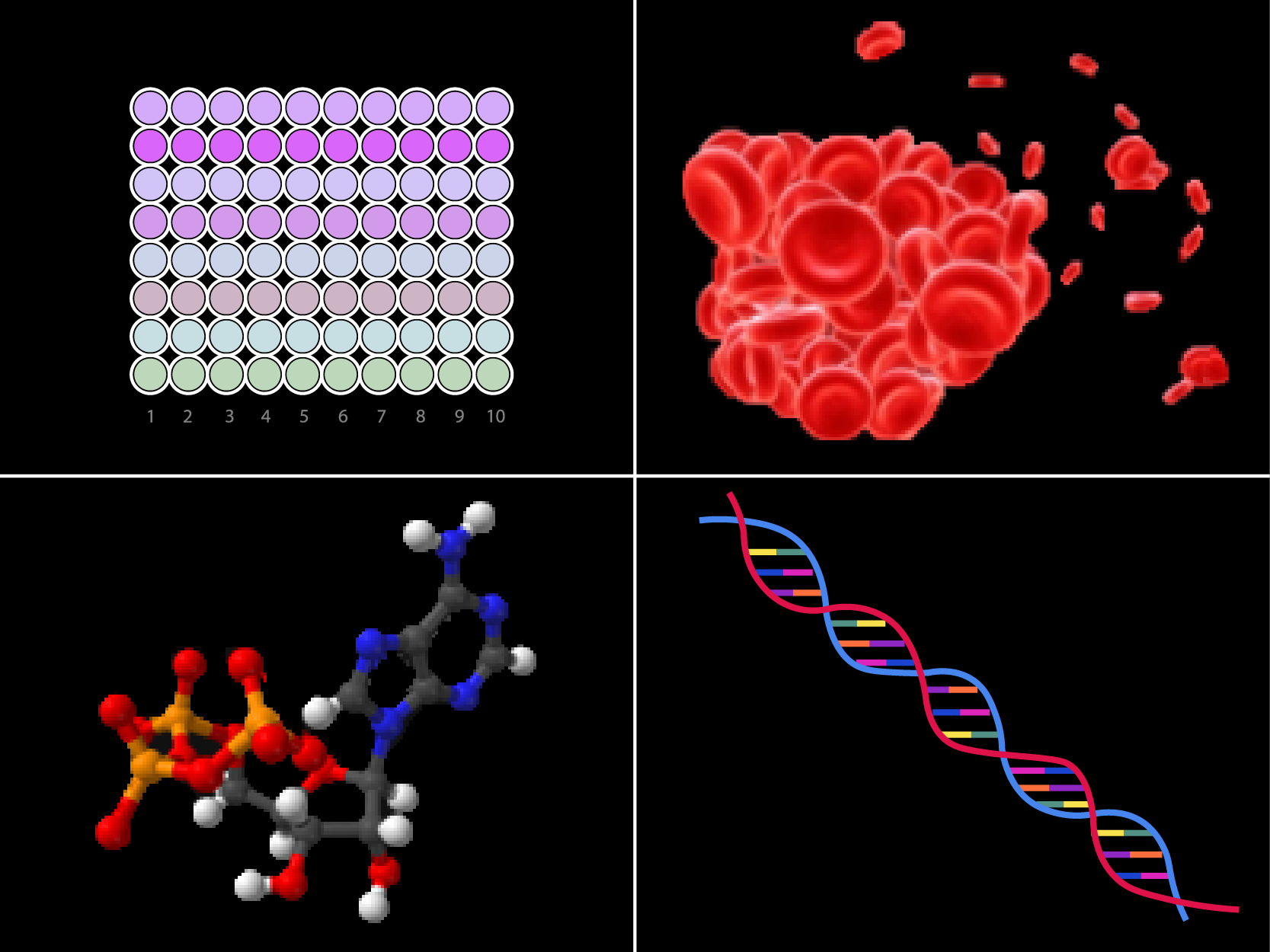
Cell proliferation assays have a wide range of applications in scientific research – from testing drug reagents to the effect of growth factors, from testing cytotoxicity to analyzing cell activity. So, what are cell proliferation assays? Cell proliferation assays typically detect changes in the number of cells in a division or changes in a cell population.
In a previous article, we explored the differences between rabbit and mouse antibodies as well as the biology behind rabbit antibody superiority. But after choosing the host, the type of technology used to produce the antibody is important too. Here, we explore some of the rabbit monoclonal antibody technologies available in the current market.
As one of the most common reagents in biology and medical research, there are more than 350,000 commercially produced antibodies available for research and clinical applications. However, the quality of the commercially available antibodies varies from vendor to vendor. Different suppliers have different protocols for validating antibodies and some researchers might want to verify the product before using them on precious samples. Here are some of the factors to examine when it comes to antibody quality.
A healthy immune system requires a series of checkpoints to ensure self tolerance and prevent damage to other tissues during immune response. Binding of costimulatory signal transduction molecules (such as CD28, ICOS, GITR) on T cells to their receptors (such as CD80/CD86, ICOSL, GITRL) on antigen presenting cells (APCs) may contribute to T cell activation. However, in some states, inhibitory signals of T cell activation and response occur during the involvement of T cell receptors. These signals are generated by proteins involved in immune checkpoints (eg, PD-1, CTLA-4, TIM-3, and LAG3). Usually PD-1 and CTLA-4 immunological checkpoint proteins are upregulated in T cells infiltrating tumors and bind to their respective ligands, PD-L1 (ligand B7-H1)/PD-L2 (ligand B7- DC) and CD80/86, and down-regulate T cell responses. Immunological checkpoint ligands are often upregulated in cancer cells as a means of evading immune detection. Therefore, immunotherapy by blocking immunological checkpoint protein activation of anti-tumor immunity has become a popular research subject for cancer therapy.
The interest in using primary cells for cell-biology research has gained prominence in recent years due to factors such as cell line contamination (Kaur G, 2012). What made primary cells lose their popularity in the first place is partly due to the rigorous and arduous process associated with primary-cell cultivation. So why is primary-cell cultivation so difficult?
The G2/M cycle checkpoint prevents cells with genomic DNA damage from entering mitosis (M phase). The main safeguards conferred by this checkpoint is to ensure that DNA is free of major lesions or replication errors, and there are enough organelles, metabolites, and other cellular cargo in the parent cell prior to division so the daughter cells can be adequately provided for once mitosis is complete. Failures at this checkpoint are associated with aberrant cellular growth and cancer progression.
The Cyclin B-CDK1 complex plays an important regulatory role during the G2 transition, at which time CDK1 is maintained inactivated by the tyrosine kinases Wee1 and Myt1. When the cells enter the M phase, the kinase Aurora A and the cofactor Bora act together to activate PLK1, which in turn activates the activity of phosphatase CDC25 and downstream CDC2, effectively driving the cells into mitosis. When the DNA is damaged, it activates the DNA-PK/ATM/ATR kinase and eventually inactivates the Cyclin B-CDK1 complex. Stopping cell cycle progression allows the cell enough time to attempt to repair any DNA or cellular damage, and if all else fails, to induce apoptosis to prevent risk to the entire organism.
ABclonal Technology provides a wide selection of cell cycle checkpoint antibody products for every phase of a cell's life. Please see a small sample of our offerings below.

