Dec 4, 2018 10:35:13 AM       by Panyue (Penny) Hao

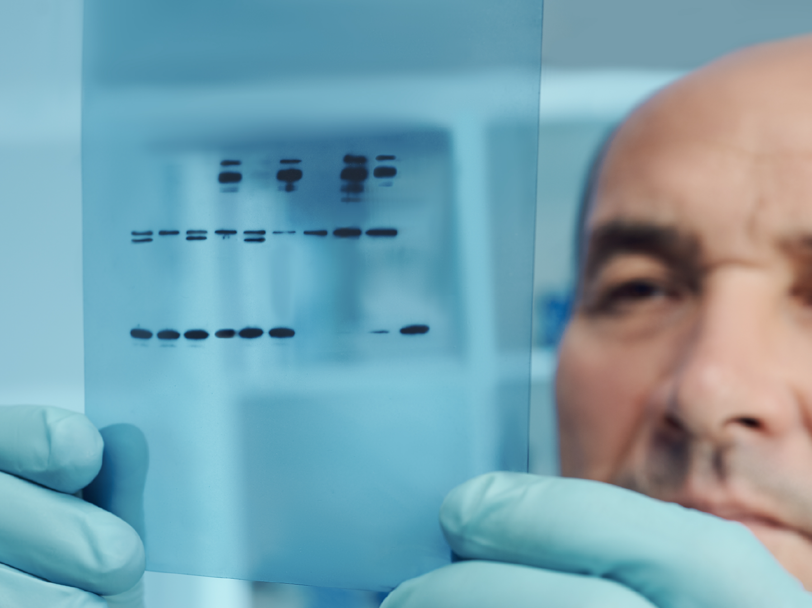
High-Dilution GAPDH Monoclonal Antibody

The glyceraldehyde-3-phosphate dehydrogenase, or GAPDH for short), is a multifunctional, indispensable enzyme found in all cells. The generally known function of GAPDH is to assist in carbohydrate metabolism as a key player in glycolysis, but there are studies demonstrating its role in the nucleus as well. 


GAPDH is a constitutively expressed housekeeping protein, and GAPDH mRNA levels and protein levels are often used as loading controls in experiments that quantify target-specific expression changes. Recent studies have elucidated the role of GAPDH in apoptosis, gene expression through its possible activities as a transcription factor, and nuclear transport. As both a metabolic protein as well as one that might play a role in cytoskeletal reorganization, GAPDH activity is intricately tied to tumorigenesis. GAPDH may also play a role in neurodegenerative diseases such as Huntington's disease and Alzheimer's disease. Therefore, although many researchers do use GAPDH as a control, this protein needs to be appreciated for its myriad other functions as well!


ABclonal Technology's GAPDH recombinant rabbit monoclonal antibody is a human-specific antibody that can be used with a high dilution ratio of 1:2560000. As a highly-stable antibody product, this means that you can perform numerous Western blotting experiments over a long period of time using a small quantity of antibody, as well as in other experiments to study the functions of GAPDH. Take advantage of this robust, cost-effective antibody product in your research today!


Nov 6, 2018 12:15:49 PM       by Panyue (Penny) Hao

Golgi Apparatus Markers

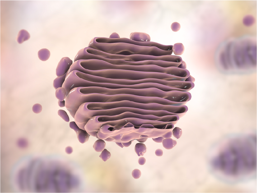
Although underappreciated, the Golgi apparatus is indispensable to normal cellular function by ensuring proteins are properly folded and sorted, and to direct diverse functions including autophagy. Disruptions to proper Golgi function can lead to many disease states, including diabetes, cancer, and neurodegenerative disorders such as Alzheimer's disease. The study of vesicular markers, including Golgi markers, is critical to our understanding of this amazing organelle's function in keeping the cell and organism healthy. Clarifying the mechanisms by which proteins are properly folded, sorted, quality controlled, and transported will prove important as more effective therapies are developed against a diverse array of human diseases.


We have previously explored the function of organelle markers USO1, GOLGA2, and GOLM1 but not how the corresponding antibodies can be applied in research. Organelle marker antibodies are common tools in cell biology research. They can be used with immunofluorescence technology to observe the morphological structure of organelles and understanding the subcellular localization of proteins. In turn, they help to explore the biological functions/role of organelle proteins in normal or disease models. These markers can also be used in Western blot (WB) experiments examining organelle extracts: as a positive control to determine whether the organelle is successfully extracted.


You can see some examples of ABclonal Technology's Golgi marker antibodies below. These are only a handful of the huge selection of targets that you can use to supplement your cutting-edge research!


Sep 25, 2018 12:20:19 PM       by Panyue (Penny) Hao

RNA Methyltransferase Antibody Featured in Cell and Nature Journals

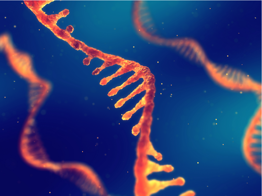
RNA methyltransferases such as METTL3, METTL14, WTAP, and VIR can catalyze the methylation of the N6 position of adenylate (M6A) and are opposed by demethylases which include FTO and ALKBH5.


Sep 11, 2018 10:58:30 PM       by Panyue (Penny) Hao

Featured Product Weekly: ER and Nuclear Membrane Markers

Endoplasmic Reticulum Marker

The endoplasmic reticulum is a membrane-bound organelle that is critical to the proper sorting and folding of proteins. Improperly folded proteins are normally allowed to refold into their functional conformation, and if not possible to repair, these unfolded proteins are directed to be degraded to prevent damage to the cell. The P4HB gene encodes a protein disulfide isomerase (PDI) that catalyzes both the formation of disulfide bonds, which form between cysteine residues to stabilize protein structure, and isomerization between or within molecules of secreted proteins. To achieve the natural conformation, this process takes place in the endoplasmic reticulum, so P4HB is often used as an ER marker. Studies on the oxidative folding mechanism indicate that molecular oxygen can oxidize the ER protein Ero1, and Ero1 can oxidize PDI through a disulfide bond. After this activity, PDI catalyzes the folding of proteins to form disulfide bonds.