CREB1 is a basic leucine zipper domain (bZIP) transcription factor that activates a target gene through a cAMP response element. As a key transcriptional regulator, CREB1 plays a role in a variety of cellular responses by mediating a number of physiological stimuli. CREB1 is expressed in many tissues and plays an especially important regulatory role in the nervous system by promoting neuronal survival, driving precursor proliferation, neurite outgrowth, neuronal differentiation and more. In addition, CREB1 signaling is involved in the learning and memory functions of many organisms. CREB1 is capable of selectively activating many downstream genes through interaction with multiple dimerization partners. Phosphorylation of CREB1 at the serine 133 site involves multiple signaling pathways, such as Erk, calcium flux (Ca2+), and stress signaling. Some of the kinases involved in CREB1 phosphorylation include p90RSK, MSK, CaMKIV, and MAPKAPK-2.
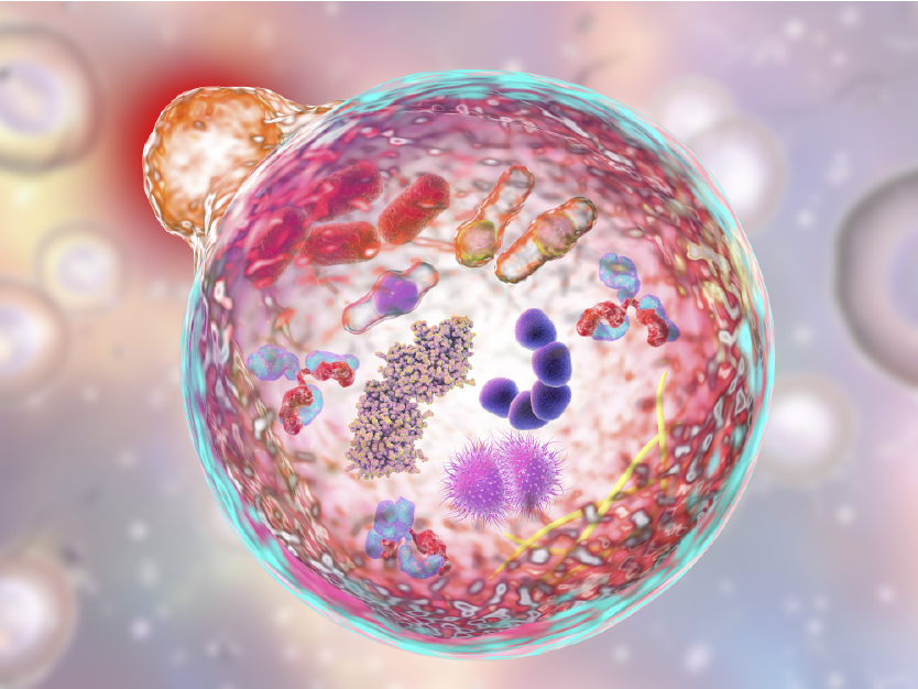
Autophagy is a catabolic process in which autophagic lysosomes, known as autophagosomes, degrade most cytoplasmic contents, including entire organelles like damaged mitochondria in protection of the host cell and organism. Autophagy is usually activated in the absence of nutrients and is associated with many physiological and pathological processes, including growth, differentiation, neurodegenerative diseases, infections and tumors. Light chain 3 (LC3) is a widely recognized autophagy marker. There are three isoforms of the LC3 protein (LC3A, LC3B, and LC3C) in mammals. They undergo post-translational modifications during autophagy. The LC3 protein is first cleaved by Atg4 at its carboxy terminus immediately after synthesis to produce LC3-I, which is localized in the cytoplasm. During autophagy, LC3-I is modified and processed by a ubiquitin-like system including Atg7 and Atg3 to produce LC3-II with a molecular weight of 14 kD and localized to autophagosomes. The magnitude of the LC3-II/I ratio can be used to assess the level of autophagy.
ABclonal Technology is pleased to offer numerous antibody products to study targets within the autophagy pathway, be it the normal functions within the pathway, the autophagic cell death pathway, or disruptions to autophagy that may drive disease progression. Many of our products have been peer reviewed by satisfied customers, including those who have used them to generate quality data for scientific publications. Please see some examples of our products below, and happy experimenting!
RNA Methyltransferase Antibody Featured in Cell and Nature Journals
RNA methyltransferases such as METTL3, METTL14, WTAP, and VIR can catalyze the methylation of the N6 position of adenylate (M6A) and are opposed by demethylases which include FTO and ALKBH5.
The nucleosome consists of an octamer composed of four histones (H2A, H2B, H3, and H4) and a DNA entangled with 147 base pairs. The core of the histones constituting the nucleosome are roughly the same, but the free N-terminus can be subjected to various modifications.
Featured Product Weekly: ER and Nuclear Membrane Markers
Endoplasmic Reticulum Marker
The endoplasmic reticulum is a membrane-bound organelle that is critical to the proper sorting and folding of proteins. Improperly folded proteins are normally allowed to refold into their functional conformation, and if not possible to repair, these unfolded proteins are directed to be degraded to prevent damage to the cell. The P4HB gene encodes a protein disulfide isomerase (PDI) that catalyzes both the formation of disulfide bonds, which form between cysteine residues to stabilize protein structure, and isomerization between or within molecules of secreted proteins. To achieve the natural conformation, this process takes place in the endoplasmic reticulum, so P4HB is often used as an ER marker. Studies on the oxidative folding mechanism indicate that molecular oxygen can oxidize the ER protein Ero1, and Ero1 can oxidize PDI through a disulfide bond. After this activity, PDI catalyzes the folding of proteins to form disulfide bonds.
The extracellular signal-regulated kinases, or ERK1/2 (MAPK1/MAPK3, p44/42MAPK), are signaling molecules belonging to the mitogen-activated protein kinase family (MAPKs) that are commonly located in the cytoplasm of eukaryotic cells. In concert with various other molecules in the signaling cascade acting under different surface or intracellular receptors, ERK1/2 act as catalysts in the phosphorylation of serine/threonine and are negatively regulated by the bispecific (Thr/Tyr) MAPK phosphatase family (called DUSP or MKP) and specific inhibitors to MEK activity (such as U0126 and PD98059).

