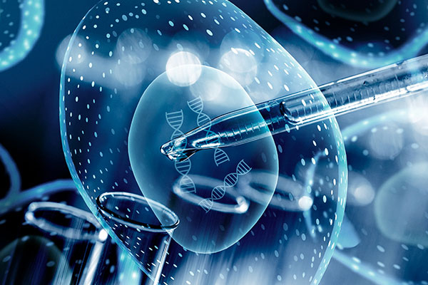
DNA methyltransferase (DNMT) is an important family of enzymes that catalyze and maintain DNA methylation, a common marker in the epigenetic silencing of target genes. The enzymes play a key role in the regulation of gene expression and genomic imprinting/development.
Featured Product Weekly: DNA Methyltransferase Antibody
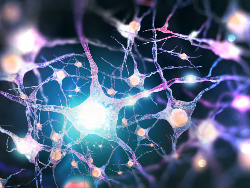
Astrocytes are specialized glial cells with distinct morphology that are found in the central nervous system, playing a role in brain and nerve cell development and the formation of synapses. Mature astrocytes respond to many stress signals and are responsible for many essential complex functions in the healthy brain, allowing the maintenance of proper homeostasis through ion flow, signaling, and the recycling of neurotransmitters. Astrocytes that are irregularly activated may result in various neurological disorders, including Alzheimer's disease and Huntington's disease.
Glial fibrillary acidic protein (GFAP) is an intermediate filament protein that is mainly found in astrocytes. It is also expressed in chondrocytes, fibroblasts, myoepithelial cells, lymphocytes, and hepatic stellate cells.
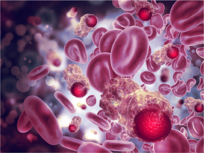
Epidermal growth factor receptor (EGFR, also known as ErbB-1 or HER1) is a member of the ErbB family. This family includes four tyrosine receptor kinases: HER1 (ErbB1, EGFR), HER2 (ErbB2, NEU), HER3 (ErbB3), and HER4 (ErbB4). The ErbB family plays an important regulatory role in the process of cell physiology, and is among the most studied receptor tyrosine kinases and signaling molecules in the history of biochemistry and cell biology.
EGFR is distributed along the surface of cells including mammalian epithelial cells, fibroblasts, glial cells, keratinocytes, and more. The EGFR signaling pathway plays an important role in physiological processes such as cell growth, proliferation and differentiation. Upon ligand binding (for example, EGF interacting with the extracellular domain of EGFR), the ErbB receptor tyrosine kinases will homodimerize or heterodimerize, allowing autophosphorylation of cytosolic tyrosine residues and the recruitment of downstream signaling molecules.
The loss of function in tyrosine kinases such as EGFR, or the abnormal activity/cell localization of key factors in related signaling pathways, such as the p38 MAPK pathway, can cause multiple cancer types, diabetes, immunodeficiency and cardiovascular diseases. In modern medicine, typical treatment strategies include targeting the tyrosine kinase activity of the ErbB receptor with small molecular inhibitors, or humanized antibodies that will target cells that have overexpressed the ErbB receptor, such as using an anti-HER2 therapy to treat breast cancer.
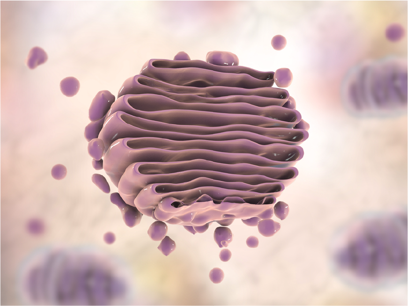
Although underappreciated, the Golgi apparatus is indispensable to normal cellular function by ensuring proteins are properly folded and sorted, and to direct diverse functions including autophagy. Disruptions to proper Golgi function can lead to many disease states, including diabetes, cancer, and neurodegenerative disorders such as Alzheimer's disease.
Part of the Golgi protein family, USO1 protein (also known as vesicle docking protein p115) is a peripheral membrane protein that can be used as a Golgi marker. It cycles between the cytoplasm and the Golgi apparatus during interphase. The position of the USO1 protein is regulated by phosphorylation -- dephosphorylated proteins bind to the Golgi membrane and dissociate from the membrane when phosphorylated. This regulated transportation plays an important role in protein localization, secretion, and signal transduction. USO1 protein acts as a vesicle anchor by interacting with the target membrane and keeping the vesicles close to the target membrane. In addition, the USO1 protein interacts with GOLGA2 (GM130) and Giantin to promote endoplasmic reticulum-Golgi transportation. A large part of Golgi-related research is in understanding how to maintain proper Golgi function to prevent and treat human diseases.
AIFM1, also known as Apoptosis Inducing Factor (AIF), is a widely expressed flavoprotein that plays an important role in caspase-independent apoptosis. AIF normally exists in the mitochondrial intermembrane space.
Nuclear lamina is a layer of cross-linked fibrin network that commonly exists in higher eukaryotic cells. It is interior to the nuclear envelope with a fiber diameter of about 10 nm. The nuclear lamina of higher animals are usually composed of three intermediate filament polypeptides – lamins A, B, and C. The nuclear lamina is closely related to the stability of nuclear envelopes, maintenance of nuclear pore location, stabilizing interphase chromatin morphology and spatial structure, chromatin construction, and nuclear assembly.

